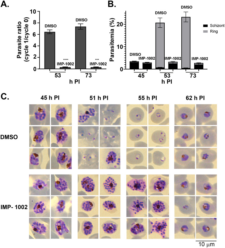
Fig 1. Inhibition of NMT during schizogony leads to a block in parasite development before merozoite egress from infected erythrocytes.
(A) The parasite ratio (parasitemia of cycle 1/ parasitemia of cycle 0) at 53 and 73 h PI measured by flow cytometry. Equal numbers of IMP-1002- and DMSO-treated parasites, collected at 45 h PI and Percoll purified, were mixed with fresh erythrocytes for a growth assay. Data are from 2 biological replicates, each in triplicate (see S1 Data). The growth of IMP-1002–treated parasites was significantly lower (p < 0.0001 [****] for 53 h PI and p < 0.0003 [***] for 73 h PI: unpaired Student t test with Welch correction not assuming an equal SD, n = 3; 50,000 RBC counted per sample). (B) Percentage of schizonts and rings in the growth assay samples. While the number of schizonts remained the same during the period from 45 h PI to 73 h PI and few rings were detected even at 73 h PI in the IMP-1002 inhibitor–treated samples, the size of the schizont population dropped and the ring population increased substantially in the DMSO-treated control culture (data are from 2 biological replicates, each in triplicate; see S1 Data). (C) Giemsa staining to reveal parasite development and morphology after 11-hour drug treatment; 6 representative parasites are displayed for each time point and treatment. While there was no visible morphological difference between IMP-1002- and DMSO-treated schizonts at 45 h PI, by 55 h PI when most parasites in the control culture were ring stages, there was an irregular distribution of merozoites within the drug-treated schizonts. Parasites that survived drug treatment developed normally into trophozoites (shown at 62 h PI) (20 fields of view per sample, n = 3). Each panel width and the scale bar are 10 μm. h PI, hours postinfection; NMT, N-myristoyl transferase; RBC, red blood cell.

