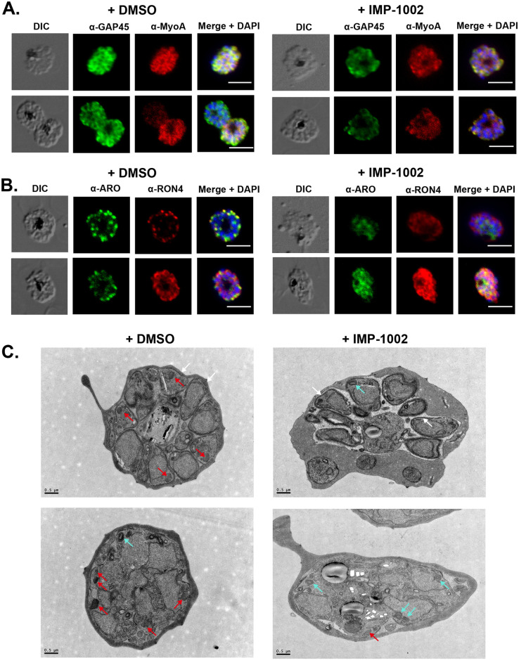
Fig 2. IMP-1002 treatment during schizogony has no effect on formation of the IMC but affects the subcellular location of ARO and RON4 and rhoptry biogenesis.
Light microscopy images from indirect IFAs performed in duplicate on 3 separate occasions with fixed parasites from DMSO (control) and IMP-1002–treated parasites and protein-specific antibodies. Panels show the DIC image, the specific antibody location (green or red), and a merged image of antibody staining with DAPI staining of nuclei. Scale bar = 5 μm. (A) Two examples showing that the location of GAP45 and MyoA at the IMC appeared largely unchanged following drug treatment. (B) Two examples showing that the location of ARO and RON4 was affected by drug treatment, with the proteins appearing to be distributed throughout the cytoplasm of developing merozoites and a loss of distinct rhoptry staining in treated parasites. (C) Transmission electron micrographs of DMSO and IMP-1002–treated schizonts. IMC appears to develop normally in both parasite populations (white arrows). Two populations of rhoptry were evident: electron-dense structures (red arrows) that were predominantly in control cells and those with a lighter core or inclusion (cyan arrows) found largely in drug-treated cells. Scale bar = 0.5 μm. ARO, armadillo domain–containing rhoptry protein; DIC, differential interference contrast; GAP45, glideosome-associated protein 45; IFA, immunofluorescence assay; IMC, inner membrane complex; MyoA, myosin A; RON4, rhoptry neck protein 4.

