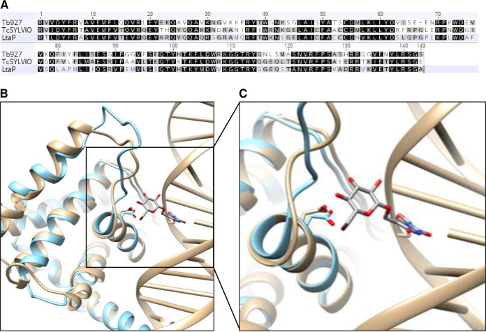
FIG 2.
Modeling of the JBP3 DNA-binding domain. (A) Sequence alignment of the putative JBP3 J-binding domains from the T. brucei EATRO927 strain, the T. cruzi Silvio strain, and the L. tarentolae Parrot strain. Residues that are identical or conservatively replaced in all three species are shaded black, while those that are identical or conserved in two species are shaded gray. (B) The structure of the DNA-binding domain from JBP3 (light blue) was modeled using RosettaCM against the J-binding domain of JBP1 (tan) from PDB accession number 2XSE. The interaction between the conserved aspartic acid residue (Asp525 in JBP1 and Asp241 in JBP3) and the glucose of base J is shown. (C) Higher-resolution view of the interaction between the conserved aspartate of both proteins and base J.

