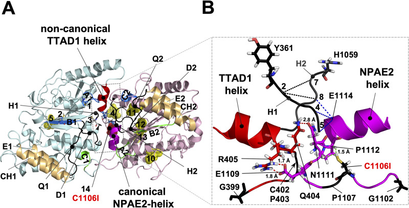
FIG 6.
Architecture of the GX2[3]CPX3NPAD/E loop-helix motifs that are in contact with each other at the cytosolic apex between the two NBDs of Cdr1. (A) Model, viewed from the top, of the nucleotide-free conformation of the NBDs of Cdr1 (24) based on the ABCG5-G8 structure (18); the TMDs were removed for clarity. The N- (turquoise) and C-terminal (pink) halves are color coded. The characteristic signature motifs of the noncanonical CNBD1 (A1, B1, C2; for abbreviations see Fig. 1) and the catalytically active canonical CNBD2 (A2, B2, C1) are shown in blue and green, respectively, and the Q-, D-, and H-loops (Q1, Q2, D1, D2, H1, and H2) are in black and indicated with lines pointing toward their center. The N-terminal (4 and 5) and C-terminal (10 to 13) cysteines are shown as yellow dots; the N-terminal cysteines 1 to 3 that are part of A1, B1, and H1, respectively, are shown as sticks; and C1106 (14) is highlighted as a red dot near the center of the converging NBDs. The coupling helices (CH1 and CH2), unique ABCG transporter features which connect TMD1 and TMD2 with the E-helices (E1 and E2) of NBD1 and NBD2, respectively, are highlighted brown. The two GX2[3]CPX3NPAD/E loop-helix motifs providing tight contact between the two NBDs at the cytosolic apex are highlighted red (NBD1; degenerate motif; TTAD1 helix) and magenta (NBD2; canonical motif; NPAE2 helix). The canonical NPAE2 helix on the edge of one half of the centrally located NBD1-NBD2 contact region is positioned right underneath the canonical CNBD2, and the noncanonical TTAD1 helix (red) on the edge of the other half of the NBD1-NBD2 contact region is positioned right underneath the noncanonical CNBD1. (B) Close-up side-on view of the peripheral contact region near the center of the NBDs delineated with dashed gray lines in panel A. To show how H1 and H2 interact closely with the conserved contact motifs underneath, the remainder of the NBDs were removed. The noncanonical H1-Y361 and the canonical H2-H1059 are shown as black sticks. The six conserved CP and G residues of the noncanonical NBD1 (red) and the canonical NBD2 (magenta) GX2[3]CPX3NPAD/E motifs are in black (C402, P403, P1107) with the two N-terminal G residues (G399 and G1102) and C1106 shown as sticks. Residues of the loop regions that are in close proximity (<3 Å) near the center of the two NBDs are also shown as sticks: Q404 is in close contact with N1111 (red dashed line), R405 forms a possible salt bridge (red dashed lines) with E1109, and C1106 is in close contact (green dashed lines) with P1112. C1106 is part of CP2, and N1111 and P1112 are part of the canonical NPAE2 helix. Close contacts (<3 Å) between H1 and H2 with E1114 of the NPAE2 motif are indicated with black (H1-H2) and blue (H1-E1114 and H2-E1114) dashed lines, respectively. The five H1 residues YQCSQ were numbered 1 to 5, and the five H2 residues HQPSAL were numbered 6 to 11, respectively. Further details of the architecture surrounding this region are provided in Fig. S5 in the supplemental material.

