
FIG 2.
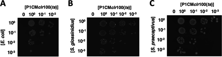
Infection of bacterial strains by phage P1. Lysates derived from an E. coli P1CMclr-100(ts) lysogen (KL463) were used to infect E. coli MG1655 (A), Sodalis glossinidius (B), and Sodalis praecaptivus (C). Plates depict the formation of chloramphenicol-resistant colonies as functions of the concentration of bacteria (vertical axis) and the concentration of P1CMclr-100(ts) lysates (horizontal axis). Note that P1 infection conditions for the strains are different (see Materials and Methods), and images do not reflect efficiency of P1 infection. Images show representative plates of at least three routine experiments.