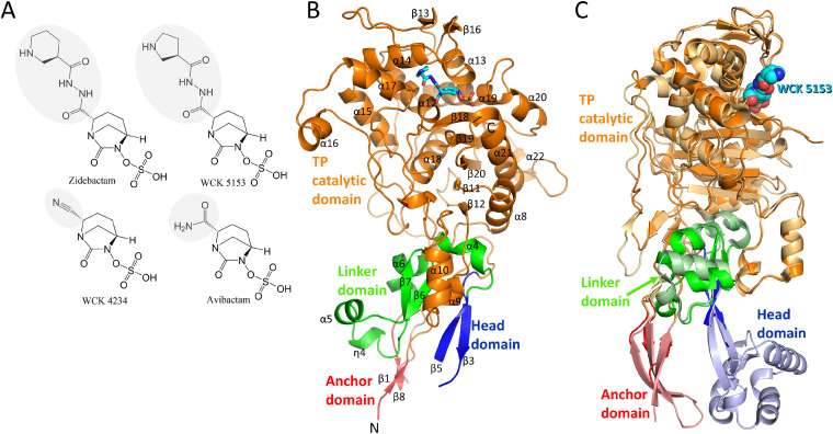
FIG 1.
Structures of DBO inhibitors and P. aeruginosa PBP2. (A) Chemical structures of DBOs zidebactam, WCK 5153, WCK 4234, and avibactam. The R1-groups of the DBOs are shaded gray. (B) Co-crystal structure of P. aeruginosa PBP2 in complex with WCK 5153 (the latter is shown in stick representation with cyan-colored carbon atoms). The PBP2 anchor (red), head (blue), linker (green), and catalytic (orange) domains are labeled, as well as most of the secondary structure elements. (C) Superpositioning of the P. aeruginosa PBP2:WCK 5153 complex and E. coli PBP2 structure. The domain color coding for P. aeruginosa PBP2 is the same as in panel B with WCK 5153 depicted in spheres with cyan-colored carbon atoms. E. coli PBP2 is shown in similar but paler colors for its respective domains.

