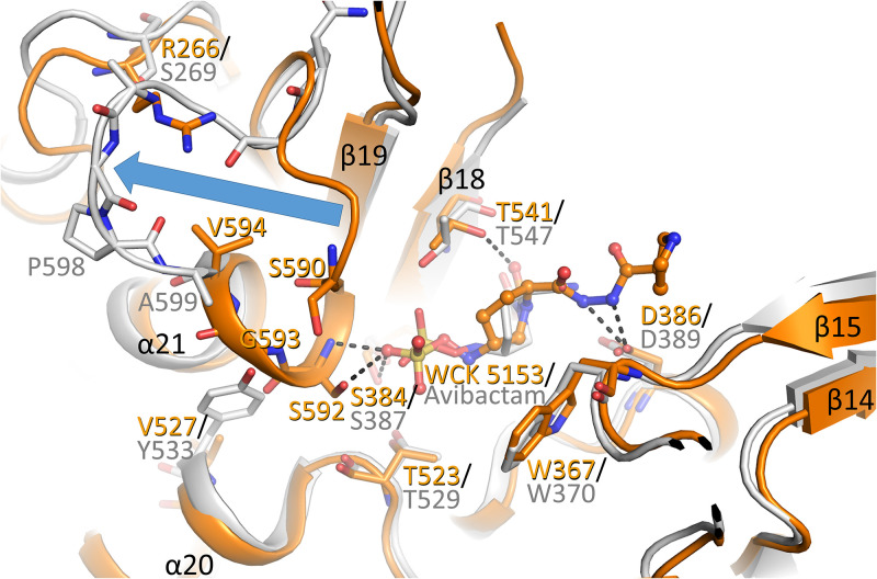
FIG 3.
Superpositioning of P. aeruginosa PBP2:WCK 5153 complex with the E. coli PBP2:avibactam complex structure. The P. aeruginosa PBP2 structure (orange) and the E. coli PBP2 structure (gray, PDB ID 6G9F) are shown with their respective ligands shown in ball-and-stick representation. The large conformational difference of the N-terminal part of the helix α21 and connecting loop in the P. aeruginosa structure from that in the E. coli PBP2 structure is depicted by the blue arrow.

