FIG 1.

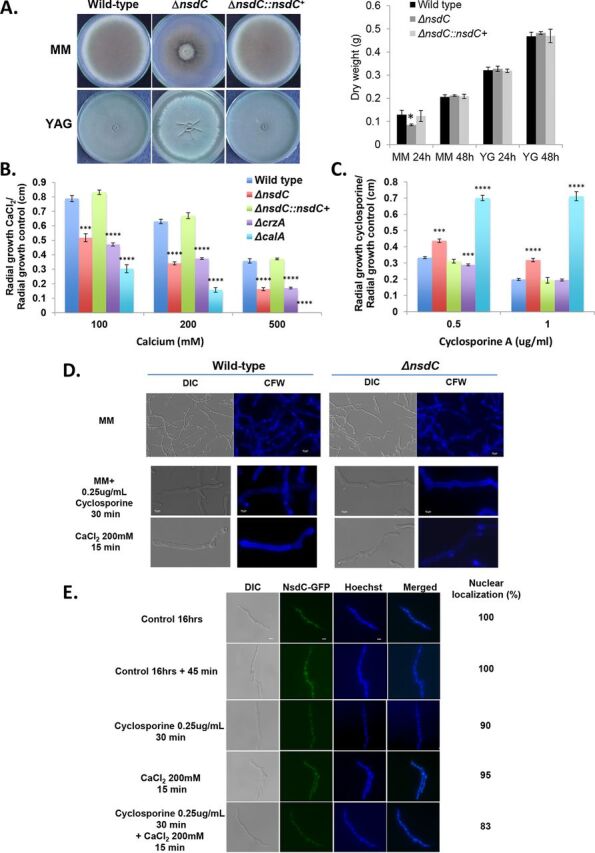
Growth phenotypes for ΔnsdC. (A to C) The wild-type, ΔnsdC, and ΔnsdC::nsdC+ strains were grown for 5 days on solid MM and YAG or for 2 days in liquid MM and YG at 37°C (A), MM + CaCl2 (B), and MM + cyclosporine (C). Results are expressed as radial growth of treatment/radial growth of control (centimeters) and are the average of three independent biological repetitions ± standard deviation. The statistical analysis was one-tailed, paired t test. P values: *, P < 0.05; **, P < 0.01; ***, P < 0.001; ****, P < 0.0001. (D) Conidia of the wild-type and ΔnsdC strains were germinated for 16 h at 37°C in liquid MM and exposed or not to 0.25 μg/ml cyclosporine for 30 min or 200 mM CaCl2 for 15 min. Germlings were stained with calcofluor white (CFW). DIC, differential interference contrast. First row shows ×40 magnification while second and third rows show ×100 magnification. (E) NsdC-GFP germlings were grown for 16 h at 37°C in MM and exposed to 200 mM CaCl2 for 15 min or 0.25 μg/ml cyclosporine for 30 min followed by 200 mM CaCl2 for 15 min. About 100 germlings were counted in each treatment. Bar, 5 μm. Statistical analysis was performed using a one-way ANOVA comparing both ΔnsdC and ΔnsdC::nsdC+ strains to the wild type (****, P < 0.0001).
