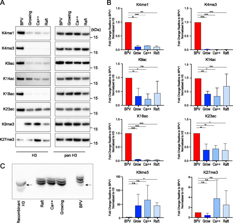
FIG 5.
BPV virions are enriched in histone H3 modifications associated with transcriptional activation and depleted for those associated with transcriptional repression. (A) The left panel shows representative immunoblots of BPV virions and bovine keratinocyte control cells using antibodies against indicated histone H3 modifications. On the right, the immunoblots were stripped and reprobed with antibody against all forms of histone H3 as a loading control. (B) Quantification of immunoblots in panel A. For each band, histone modification signals were normalized to the corresponding bands from the panH3 signal. The resulting levels are represented relative to the signal for BPV1 (K18ac, K4me1, K4me3, K9me3; n = 2) (K9ac, K14ac, K23ac, K27me3; n = 3). Error = SD. Significance was determined by an unpaired t test. n.s., P > 0.05; *, P < 0.05; **, P < 0.01; ***, P < 0.001; ****, P < 0.0001. (C) PanH3 immunoblot against an acid-urea separation of histones from BPV1 virions and bovine cellular controls. The more slowly migrating, upper bands correspond to an increase in acetylation of H3 lysine residues. Recombinant H3 protein (lane 1) indicates the positions of unmodified histone H3. n = 2.

