
FIG 3.
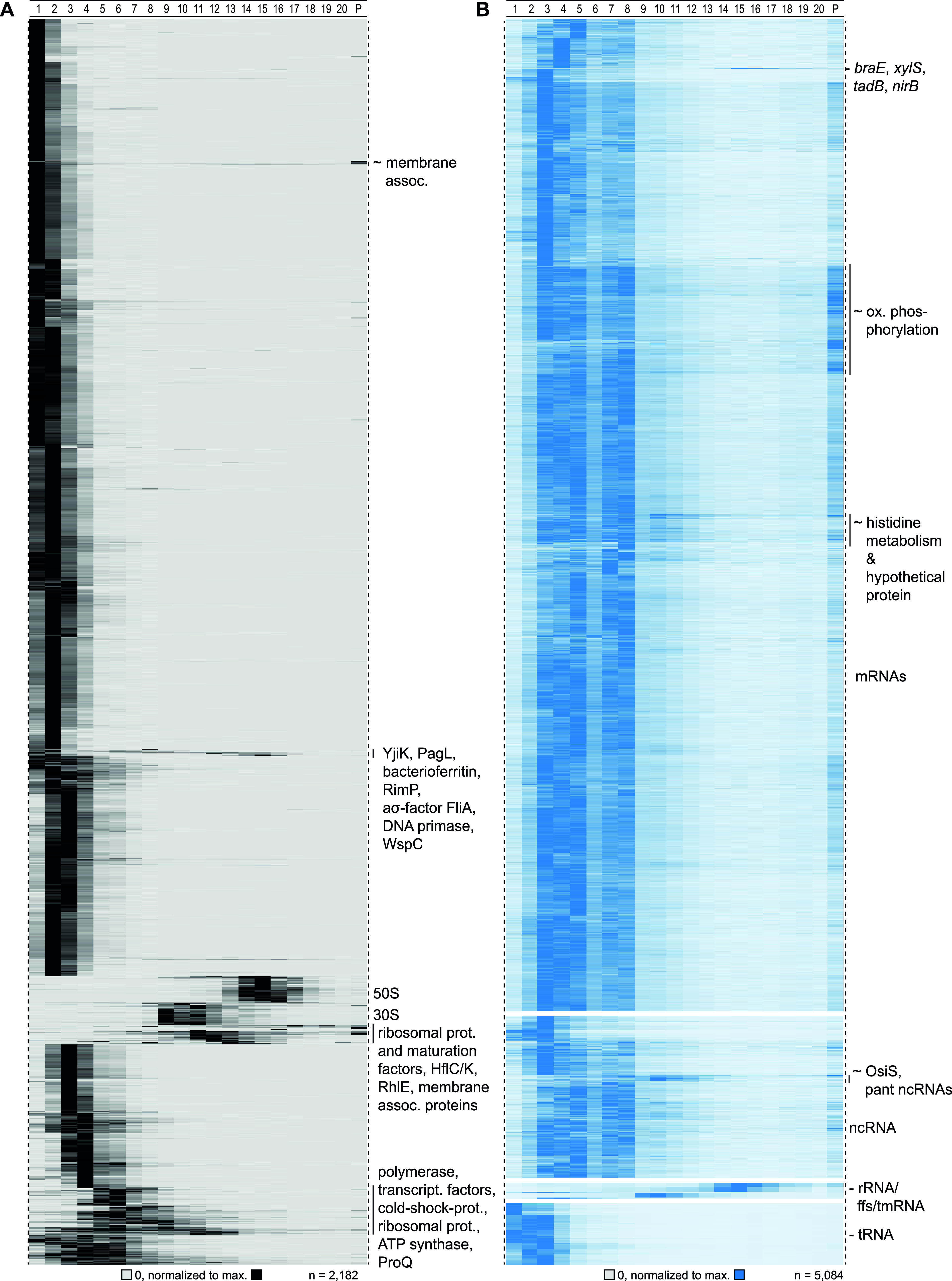
Detected sedimentation profiles in Grad-seq. (A) Proteins sedimented mainly in the first couple of fractions. Defined clusters appeared in fraction below 10. Ribosomal proteins sedimented around fractions 10 and 16. (B) Transcripts (merged CDS and UTRs) sedimented mainly in RNAP and ribosomal fractions. tRNAs did not enter the gradient, and ribosomal RNAs were allocated to HMW fractions.