Fig. 2.
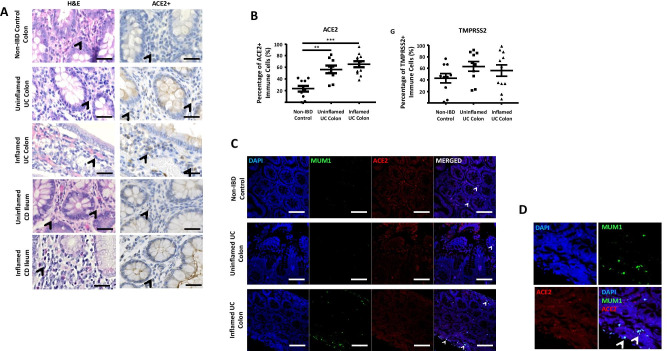
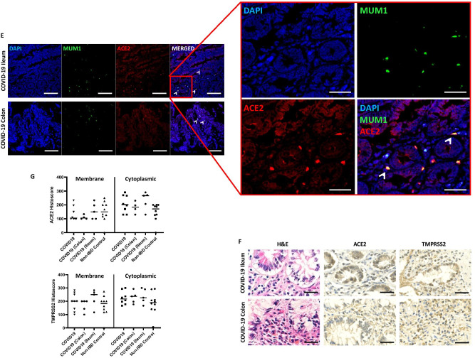
A H&E and immunohistochemistry of ACE2 in non-IBD (n = 10), UC (all colon) and CD (ileum and colon) (n = 11) patients. Representative images of non-inflamed and inflamed colonic and ileal mucosal sections. Arrowhead indicating plasma cells. B ACE2 and TMPRSS2 positive cells (%) within lamina propria in inflamed and uninflamed colon of UC vs. non-IBD controls (**p < 0.001 and *** p < 0.0001). C, D Immunofluorescence co-staining of ACE2 (Red), MUM1 (Green), and DAPI (Blue) as nuclear stain within the lamina propria of in inflamed and uninflamed colon of UC vs. non-IBD controls. E Immunofluorescence co-staining of ACE2 (Red), MUM1 (Green) and DAPI (Blue) as nuclear stain within the lamina propria of expression in fatal COVID-19 (SARS-CoV-2 PCR positive) ileum and colon sections; and non-IBD controls. F H&E and immunohistochemistry of ACE2 and TMPRSS2 protein expression in fatal COVID-19 (COVID-19 colon and ileum — n = 4 and 5 mucosal sections respectively). G ACE2 and TMPRSS2 protein weighted histoscore expressions within membrane and cytoplasm of intestinal enterocyte (COVID-19 colon and ileum — n = 4 and 5 mucosal sections respectively and 9 non-IBD colon).


