. 2021 Oct 12;10:e70910. doi: 10.7554/eLife.70910

Figure 6. Genes that promote extraintestinal pathogenic Escherichia coli (ExPEC) survival in the liver.

(A) Inverse volcano plot of fitness of ExPEC genes in the liver. Genes shown in red are involved in cell envelope homeostasis, and genes shown in blue are involved in generating fructose-6-phosphate, and their roles are schematized in (B). (B) Pathways to convert sugars to fructose-6-phosphate. GlcNAc generated from peptidoglycan (PG) degradation is converted to GlcNaC-6P by nagK, which is not among the underrepresented genes in (A).

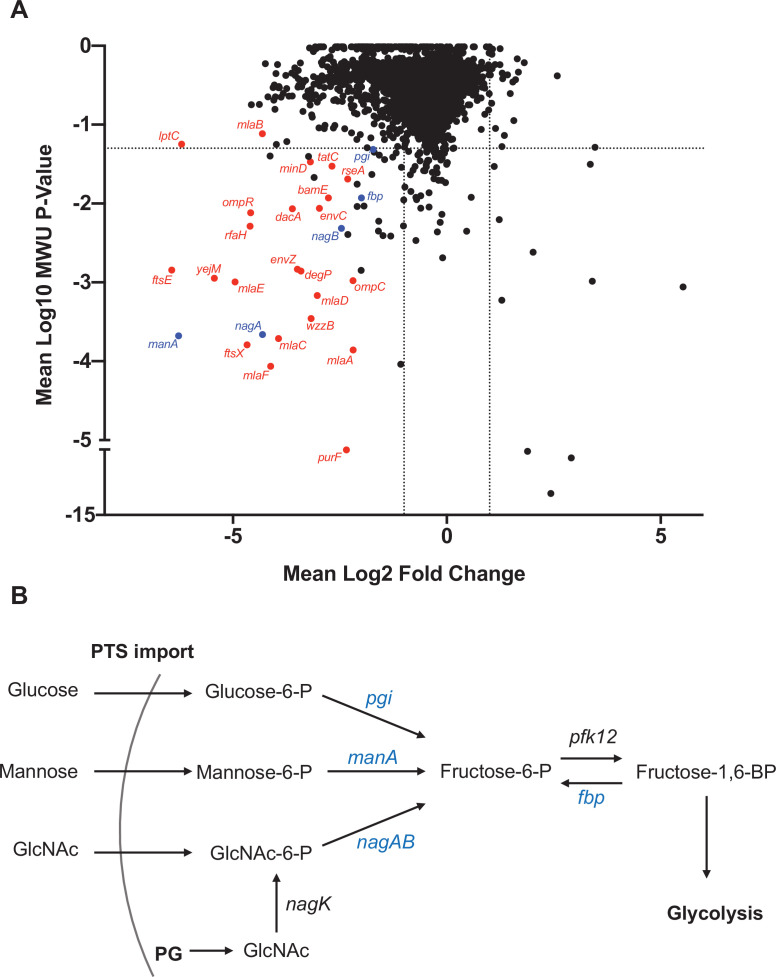
Figure 6.

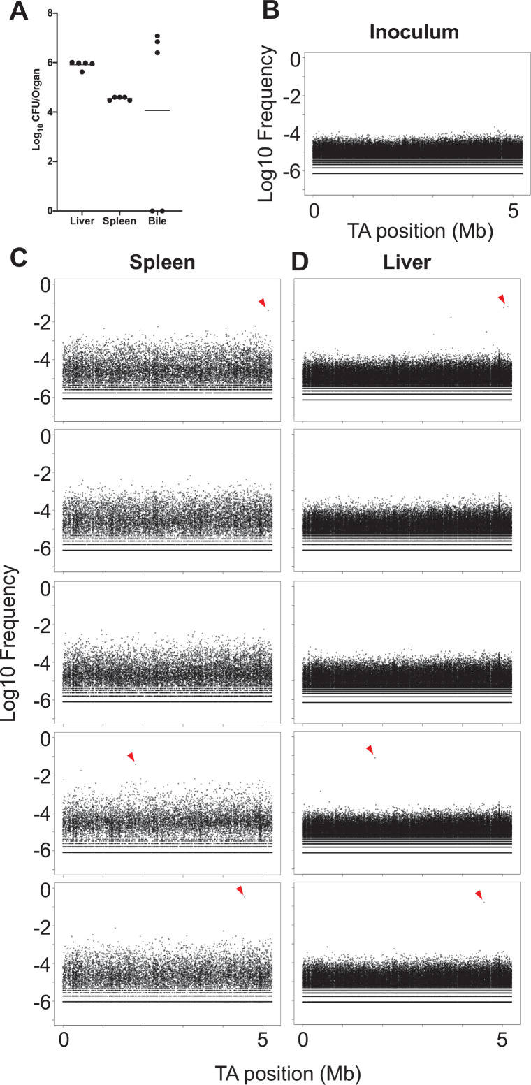
Figure 6—figure supplement 1. CFU and population structure from in vivo transposon mutant screen.

Figure 6—figure supplement 1.

(A) Bacterial burdens for the liver, spleen, and bile are shown for animals sacrificed 1 day post infection (dpi) following inoculation of the transposon library. The three animals with bile CFU correspond to the three animals where spreading of clones between the liver and spleen was observed (C, D). (B–D) Frequencies of mutants (analogous to barcodes) are shown as a function of genome position. Mutants are relatively evenly represented in the inoculum (B). (C, D) Distributions from the livers and spleens from five animals. Three animals possessed marked expansion of a single mutant that spread between the liver and spleen (red triangles).