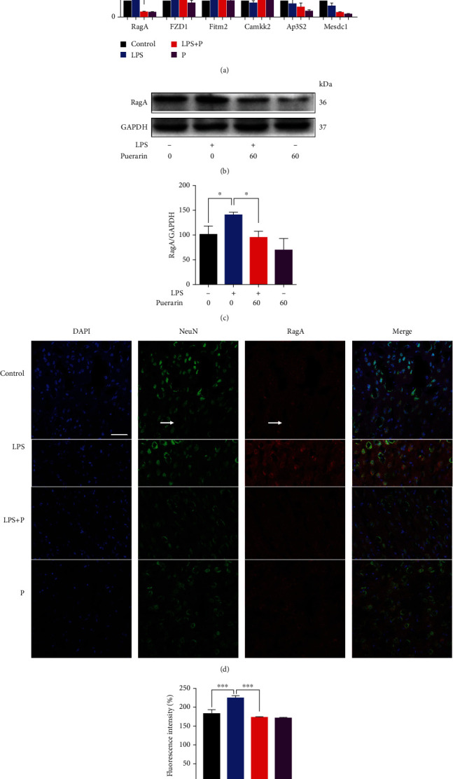
Figure 3.

Puerarin downregulated RagA expression in LPS-challenged mice. (a) qRT-PCR quantification of 6 representative differentially expressed genes. The frontal cortex tissues were collected from 4 groups of mice (i.e., control, LPS, LPS+puerarin (60 mg/kg), and puerarin (60 mg/kg), n = 3) and analysed by qPCR for selected genes. The data were presented as means ± SEM (n = 3). ∗p < 0.05. (b) Western blot analysis of the protein expression of RagA. The frontal cortex tissues were collected from the experimental mice (n = 3) for Western blot analysis of RagA. (c) Quantification of RagA expression. The blots were detected by a densitometric method (n = 3). ∗p < 0.05. (d) Immunofluorescence detection of RagA expression in the frontal cortex. The frontal cortex tissues were collected from the experimental mice (n = 3) for immunohistochemical analysis for RagA expression, whereas DAPI was used to stain nuclear. NeuN was detected as the biomarker for the mature neurons. The images were captured with a Carl Zeiss 700 confocal fluorescence microscope (Jena, Germany). Scale bar, 50 μm. (e) Quantification of RagA expression. Fluorescence intensity of RagA in (d) was evaluated through the densitometric method (n = 3). ∗∗∗p < 0.001.
