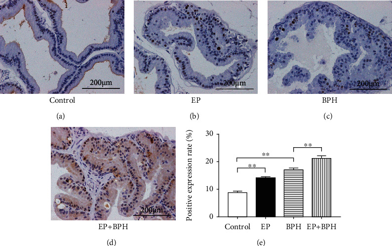
Figure 3.

Ki67 staining for detecting cell proliferation of prostate tissues. (a–d) Representative figures from Ki67 immunohistochemical staining for ventral prostates of the control, EP, BPH, and EP+BPH groups, respectively. Scale bar = 200 μm; original magnification ×400. (e) A bar graph for rate (%) of Ki67-positive cells in rat prostate. Data are presented as mean ± SEM (∗∗p < 0.01).
