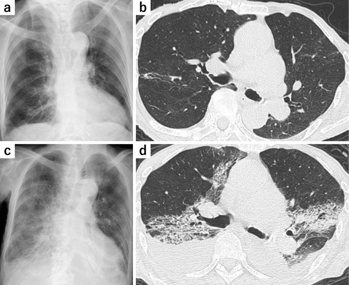
Figure 1.
Radiological findings. Chest X-ray (a) and chest CT (b) on admission showing some old inflammatory changes but no findings of active inflammation. Chest radiograph (c) and chest CT (d) on day 6 post-admission showing bilateral symmetric ground-glass opacities associated with consolidations and bilateral pleural effusions. CT: computed tomography

