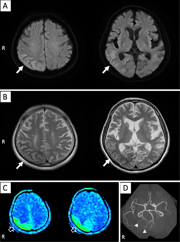
Figure 1.
Magnetic resonance imaging findings at admission. (A) Diffusion-weighted imaging and (B) T2-weighted imaging revealed cortical high intensity and subcortical low-intensity signals on the right parietal lobe, adjacent upper segment of the right temporal lobe, and lateral aspect of the right occipital lobe (solid white arrows). (C) Arterial spin labeling showed high signals in the same area where the abnormal findings were observed on magnetic resonance imaging (open white arrows). (D) Magnetic resonance angiography revealed an increased signal intensity in the right middle and posterior cerebral arteries (solid white arrowheads).

