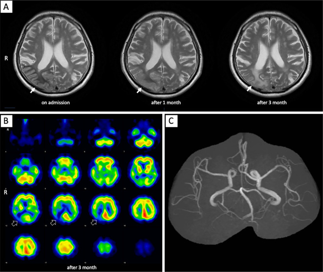
Figure 3.
(A) Subcortical low-intensity signals in the right parieto-occipital lobe on T2-weighted imaging had become an iso-intensity signal at one month after admission (solid white arrow). Subcortical abnormal signals in the right parieto-occipital lobe had become a high-intensity signal at three months after admission. (B) 123I-IMP brain perfusion single-photon emission computed tomography revealed a decreased accumulation at the right parieto-occipital lobe (open white arrows). (C) The dilated right middle and posterior cerebral arteries returned to normal on magnetic resonance angiography.

