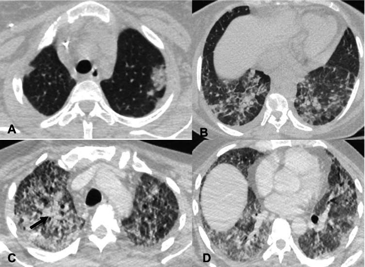
Fig. 12.
A and B—Initial axial non-contrast chest CT images in a 68-year-old man show ground glass opacities in the left upper lobe and bilateral lower lobes concerning for COVID-19 pneumonia. The patient subsequently developed COVID-19 ARDS and was supported with mechanical ventilation. Axial contrast-enhanced CT images (C and D) were obtained two months later when the patient had increasing secretions and worsening fever. There are new consolidations in the right upper lobe (arrow) and worsening ground glass opacities in the lower lobes. Cultures from the endotracheal tube secretions were positive for Klebsiella pneumoniae and Streptococcus pneumoniae, consistent with ventilator associated pneumonia (VAP)

