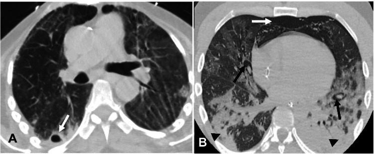
Fig. 8.
A—Lung tension cysts. A—Axial non-contrast chest CT image in a 78-year-old man with COVID-19 pneumonia on mechanical ventilation shows cavitary lesion/lung tension cyst in the subpleural right lower lobe (arrow). B—Pulmonary interstitial emphysema. Non-contrast axial chest CT image in a 45-year-old man with COVID-19 ARDS on mechanical ventilation shows pulmonary interstitial emphysema (PIE). Note the linear lucency surrounding bronchi/ pulmonary artery which is consistent with PIE (black arrows). Air is seen anteriorly consistent with pneumomediastinum (white arrow). Also seen are dependent consolidative opacities in the posterior lower lobes consistent with COVID-19 ARDS (arrowheads)

