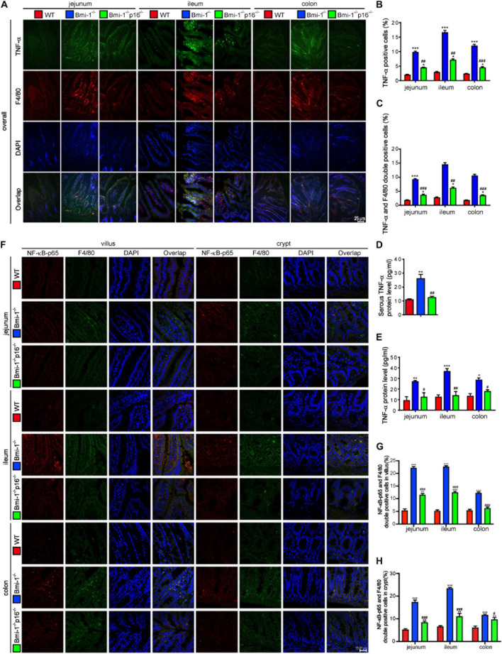
FIGURE 5.
P16 deletion improved TNF-α–dependent epithelial barrier destruction in Bmi-1–/– mice. The experiments were carried out on the 7-week-old Bmi-1–/–, Bmi-1–/–p16–/–, and WT mice. (A) Representative micrographs showing immunofluorescence for TNF-α and F4/80, with DAPI staining the nuclei. (B) Percentage of TNF-α–positive cells. (C) Percentage of TNF-α and F4/80 double-positive cells. (D) Serous TNF-α protein level (pg/mL) was detected with ELISA assay. (E) TNF-α protein levels in jejunum, ileum, and colon (pg/mL) were detected with ELISA assay. (F) Representative micrographs showing immunofluorescence for NF-κB–p65 and F4/80, with DAPI staining the nuclei. (G,H) Percentage of NF-κB–p65 and F4/80 double-positive cells or areas in villus and crypt. Six mice per group were used for experiments. Statistical analysis was performed with one-way ANOVA test. Values are mean ± SEM from six determinations per group, *p < 0.05, **p < 0.01, ***p < 0.001 compared with the WT group; #p < 0.05, ##p < 0.01, ###p < 0.001 compared with the Bmi-1–/– group.

