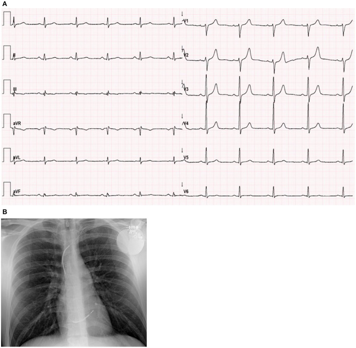
Figure 1.
Electrocardiogram and Chest X-ray. (A) ECG showed sinus rhythm, 60 bpm, normal axis, fragmented QRS in the inferior leads (III, aVF) and flat T waves in III, aVF and V6. When compared to previous ECGs, no new abnormalities were observed. (B) Chest X-ray with a single lead ICD, normal heart size and no signs of pulmonary congestion.

