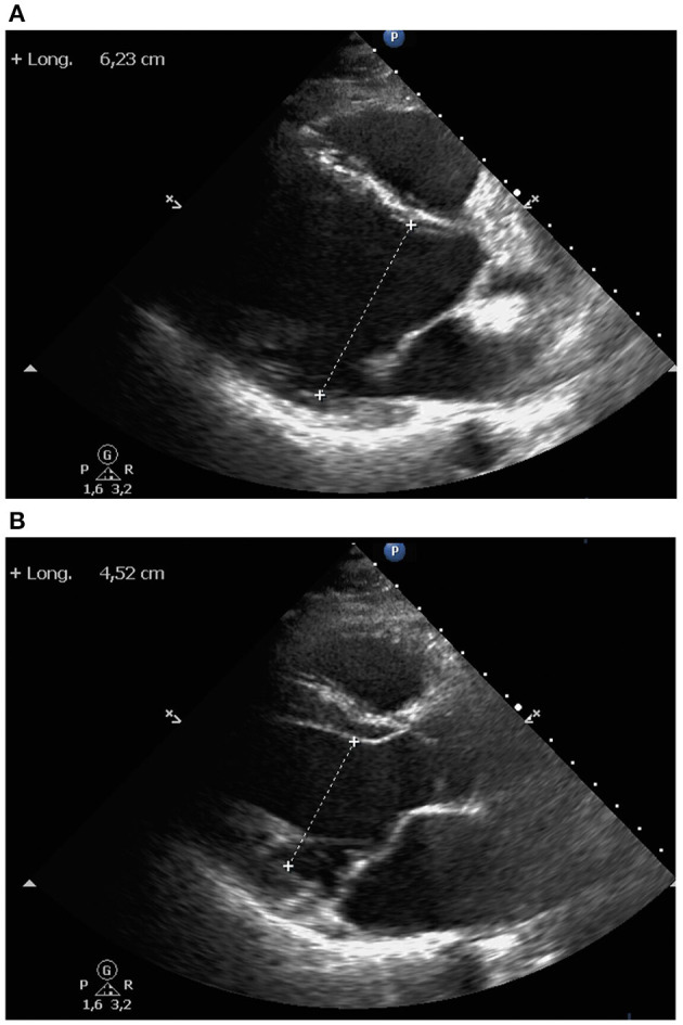
Figure 2.

Echocardiography. Parasternal long axis view showed mild left ventricular enlargement, both left ventricular end-diastolic (A) and end-systolic (B) diameters. Right ventricle, ascending aorta and left atrium were of normal size. Interventricular septum and posterior left ventricular wall had normal thickness. No pericardial effusion was observed.
