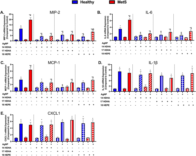
Fig. 4.
AgNP-induced pulmonary inflammatory gene expression including (A) macrophage inflammatory protein-2 (MIP-2), (B) interleukin-6 (IL-6), (C) monocyte chemoattractant protein-1 (MCP-1), (D) interleukin-1β (IL-1β), and (E) chemokine 1 (CXCL1) were evaluated in healthy and MetS lung tissue. Thirty minutes prior to oropharyngeal aspiration of pharmaceutical grade sterile water (control) or AgNPs (50 μg) in sterile water, mice were i.p. injected with 1μg of 14-HDHA, 17-HDHA, or 18-HEPE or vehicle (250 μl of sterile saline). Values are expressed as mean ± SEM (n = 6–8/group). * denotes significant differences due to AgNP exposure comparing to model-matched control receiving the same treatment, # denotes significant differences between healthy and MetS mouse models receiving the same treatments and exposure, $ denotes significant differences due to lipid interventions comparing to groups not receiving treatment but the same exposure (p < 0.05).

