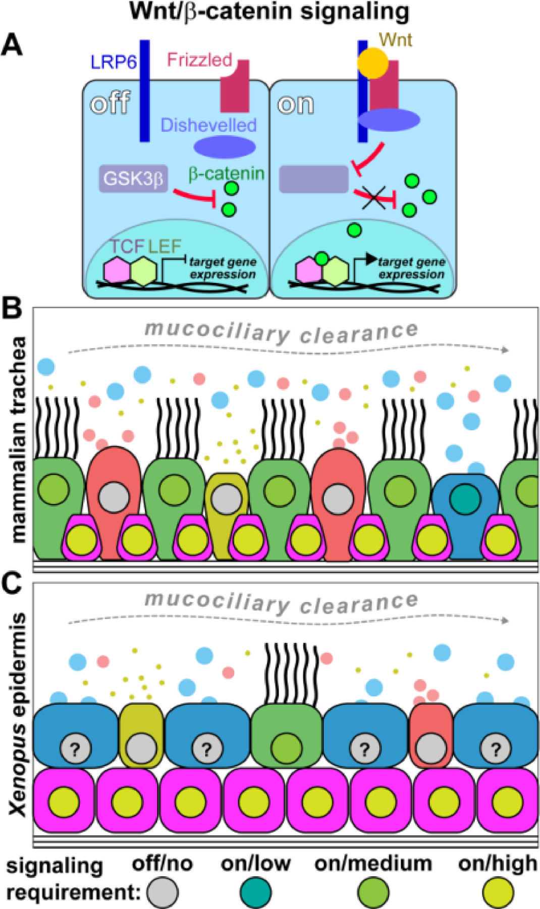
Figure 2: Wnt/β-catenin signaling requirements in mucociliary cell types.

(A) Simplified schematic representation of the canonical Wnt/β-catenin pathway. Wnt ligands bind to Frizzled receptor and recruit LRP6 co-receptor. Intracellular mediators, e.g. Dishevelled, are recruited to the ligand-receptor complex, which then leads to inhibition of GSK3β and the stabilization of β-catenin. β-catenin can then enter the nucleus, bind to TCF/LEF transcription factors, and activate Wnt target gene expression. (B) Schematic representation of cell types in the mammalian trachea and their proposed requirement levels for active Wnt/β-catenin signaling. Basal cells need high Wnt levels, while MCCs need elevated Wnt but at lower levels than basal cells. Goblet cells also require Wnt, but at low levels that heva to be precisely controlled. (C) Schematic representation of cell types in the Xenopus embryonic mucociliary epidermis and their proposed requirement levels for active Wnt/β-catenin signaling. Basal cells require high Wnt levels, while MCCs also show elevated Wnt activation. Ionocytes and small secterory cells do not seem to strictly rely on Wnt signaling, and the role for Wnt in goblet cells remains unresolved.
