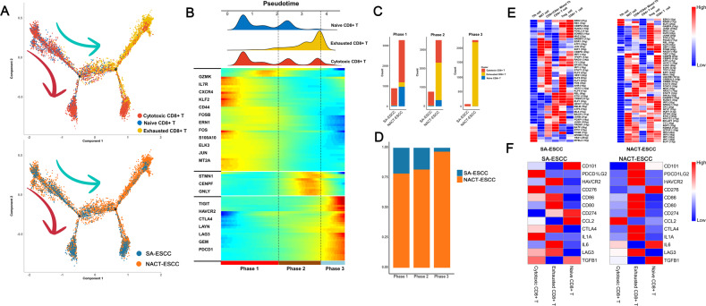
Fig. 8. The scRNA Profiles for CD8+ T cells in SA-ESCC and NACT-ESCC.
A Potential developmental trajectory of CD8+ T cells inferred by analysis with Monocle2. B Dynamic changes in gene expression of CD8+ T cells during the transition (divided into 3 phases), subtypes are labeled by colors (upper panel). C Histogram showing the cell distribution of CD8+ T cells, in SA-ESCC and NACT-ESCC samples. CD8 subtypes labeled by colors. D Histogram showing the cell distribution of SA-ESCC and NACT-ESCC samples. E Heatmap showing the activity of TFs in each CD8+ T cell subtypes in each condition. The TF activity is scored using AUCell. Left, SA-ESCC. Right, NACT-ESCC. F Heatmap showing the activity of immune checkpoints in each T cell subtypes in each condition. Left, SA-ESCC. Right, NACT-ESCC.

