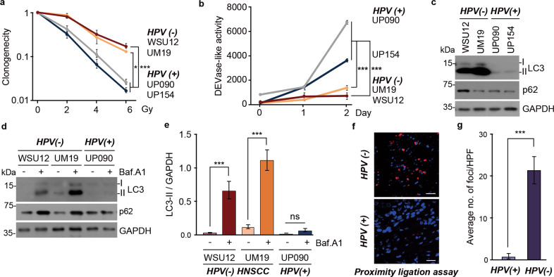
Fig. 1. Defective apoptosis and increased autophagic flux in radioresistant HPV(-) HNSCC.
a Clonogenic cell survival assay of HPV( + ) HNSCC cell lines (UP090 and UP154) and HPV(-) HNSCC (WSU12 and UM19) upon ionizing radiation (XRT) (*p < 0.05, ***p < 0.001). b DEVDase (caspase-3/caspase-7) activity assay in HNSCC cells at indicated time points post irradiation at 6 Gy (***p < 0.001). c Immunoblotting analysis of LC3 or GAPDH in HPV(-) HNSCC cell lines (WSU12 and UM19) and HPV( + ) HNSCC cell lines (UP090 and UP154). d Autophagic flux assay using WSU12, UM19 or UP154 cells in the absence or presence of bafilomycin A (500 nM, 6 h). e Quantification of LC3-II band intensity of d. Error bars represent means ± SD from three independent experiments (***p < 0.001, ns: non-significant). f In situ detection of autophagosomes in HPV(-) and HPV( + ) HNSCC tissues by p62 and LC3 PLA. Scale bar: 30 μm. g The average number of positive foci in p62 and LC3 PLA quantitated at 10X magnification (n = 4, ***p < 0.001).

