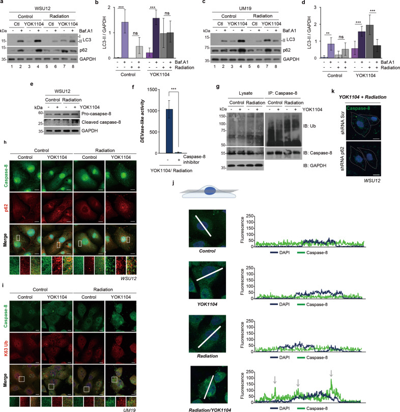
Fig. 5. YOK1104-activated p62 sequesters ubiquitinated caspase-8 into aggresome-like structures in response to irradiation.
a Autophagic flux assay using WSU12 cells treated with 5 μM YOK1104 (12 h) and/or 500 nM bafilomycin A1 (6 h) with or without irradiation at 6 Gy. b Quantification of LC3-II band intensity of a. Error bars represent means ± SD from three independent experiments (***p < 0.001, ns non-significant). c Autophagic flux assay using UM19 cells treated with 5 μM YOK1104 (12 h) and/or 500 nM bafilomycin A1 (6 h) with or without irradiation at 6 Gy. d Quantification of LC3-II band intensity of c. Error bars represent means ± SD from three independent experiments (**p < 0.01, ***p < 0.001, ns non-significant). e Immunoblot analysis of caspase-8 of WSU12 cells in the presence or absence of 5 μM YOK1104 with or without irradiation at 6 Gy. f Caspase-3/7 (DEVDase)-like activity was measured in WSU12 cells treated with 5 μM YOK1104 and 6 Gy XRT in the absence or presence of caspase-8 inhibitor (20 μM, 25 h). ***p < 0.001. g Immunoblot analyses of ubiquitinated proteins and caspase-8 using total WSU12 cell lysates (left panel) or caspase-8 immunoprecipitates (right panel) after treatment with 5 μM YOK1104 with or without irradiation at 6 Gy. h Immunostaining of caspase-8 (green) and p62 (red) in WSU12 HPV(-) HNSCC treated with vehicle control (DMSO) or 5 μM YOK1104 with or without irradiation at 6 Gy. Scale bar: 10 μm. i Immunostaining of caspase-8 (green) and K63-linked ubiquitin (red) in UM19 HPV(-) HNSCC treated with vehicle control (DMSO) or 5 μM YOK1104 with or without irradiation at 6 Gy. Scale bar: 10 μm. j Analysis of subcellular localization of caspase-8 (green) using a single focal plane at the middle of the nucleus (DAPI, blue). k Immunostaining of caspase-8 (green) in control and p62 KD WSU12 HPV(-) HNSCC treated with 5 μM YOK1104 and irradiation at 6 Gy. Scale bar: 10 μm.

