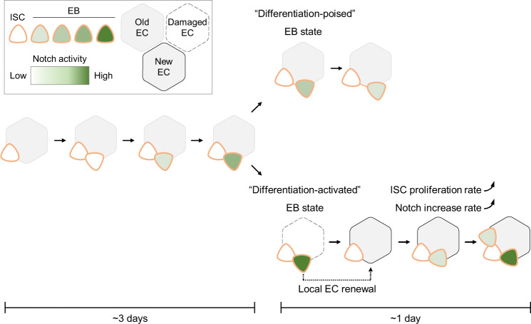
Fig. 7. Model of physiological dependence of EB differentiation in the Drosophila intestines.
The figures from the left side to the right, show an ISC that is divided once generating one self-renewal ISC and one EB with the activated Notch signaling. Notch signaling gradually increased in this EB in the intestines of young and healthy flies. This process could take more than 3 days. Later on, there were two distinct EB statuses depending on the local physiology: (1) A “differentiation-poised” EB status, corresponding to no EC damage nearby. Notch activation in this “differentiation-poised” EB could be even decreased due to the detachment from the ISC. (2) A “differentiation-activated” EB status, corresponding to a damaged EC nearby. ISC proliferation and Notch activation in EBs were dramatically increased. The local EC renew could be thus achieved in less than one day.

