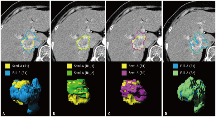
Fig. 3. Illustration of intraobserver and interobserver reproducibility of lesion segmentation.
A 66-year-old female with cholangiocarcinoma showing arterial phase hyperenhancement, who did not show recurrence during 83.8 months follow-up after surgical resection.
A. Yellow is representative of semi-automatic segmentation and blue is representative of fully automatic segmentation, by radiologist 1.
B. Light green is representative of the second trial of semi-automatic segmentation by radiologist 1. C. Purple is representative of semi-automatic segmentation by another radiologist, radiologist 2. D. Green is representative of fully automatic segmentation by radiologist 2.

