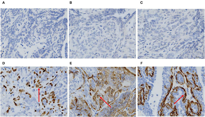
Figure 2.
Immunohistochemical staining of the primary carcinoma sections (400X). The dark brown color was observed as positive staining. Tissues from the primary cancer were negative for ER (A), PR (B) and HER-2 (C), and were positive for P63 (D), CK5/6 (E), and α-SMA (F) localized in the cell membrane.

