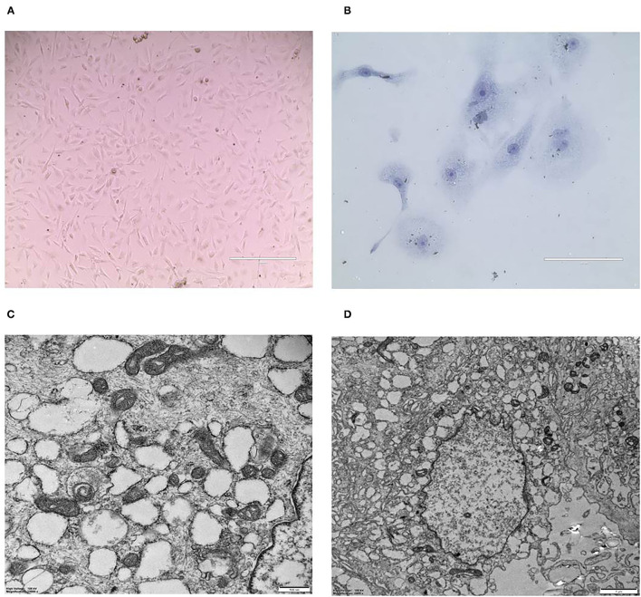
Figure 3.
Morphology of CMT-1026 cells. (A) Purified CMT-1026 cells, detected with inverted microscopy (10×). (B) CMT-1026 pellet; Diff-Quik staining (40×). Malignant cancer cells with epithelial morphology, marked anisokaryosis, and multinucleated giant cells. (C) Ultrastructural experiments revealed that CMT-1026 contained multiple organelles, mitochondria, secretory vacuoles, and endoplasmic reticulum. (D) Degenerated CMT-1026 cells, showing shrunken nuclei, and abundant proteinaceous secretion.

