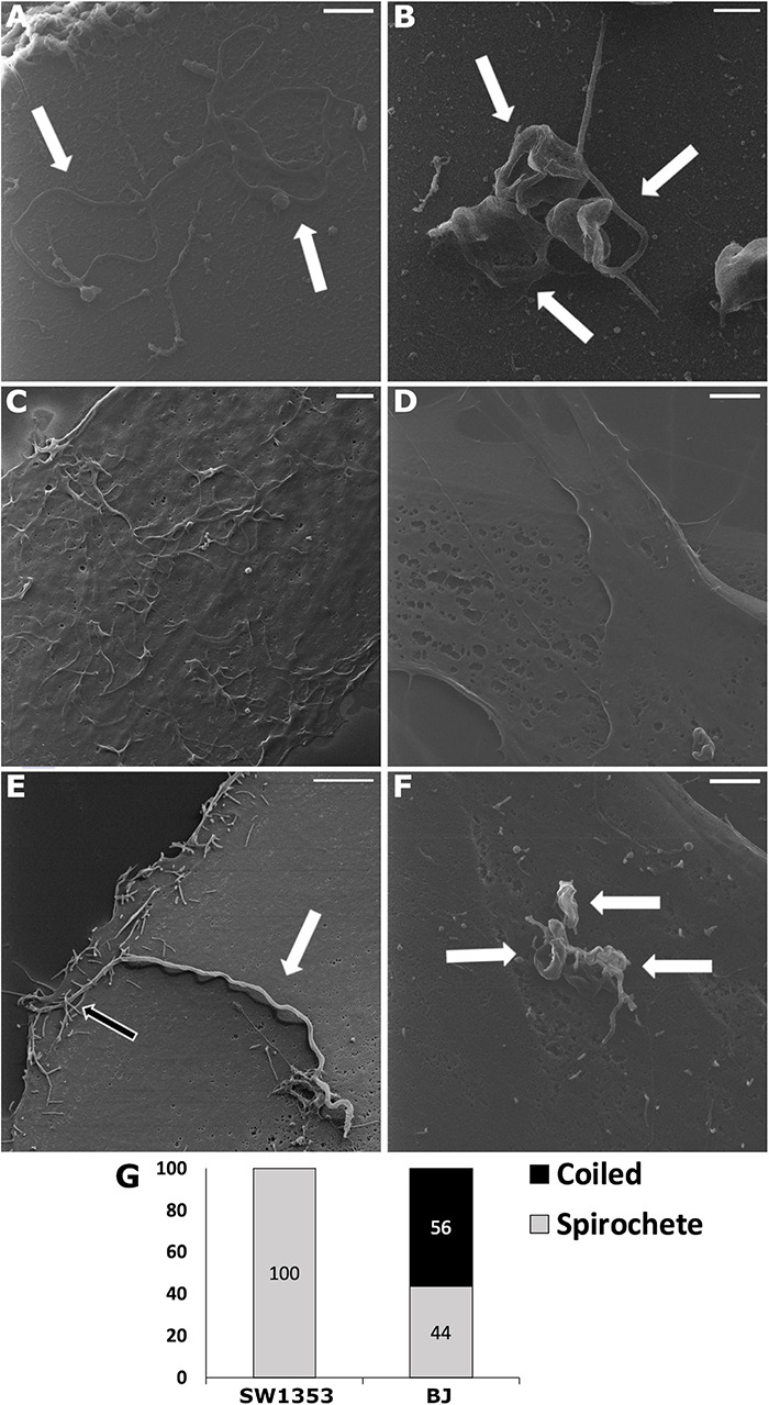
FIGURE 1.

Upon infection, human cell lines demonstrated Borrelia burgdorferi forms differently. Helium ion microscopy (Zeiss Orion Nanofab) images of Borrelia spirochetes (A) and round bodies (B), as well as, uninfected chondrosarcoma (SW1353) (C) and dermal fibroblast (BJ) (D) cells. (E,F) Borrelia spirochetes and coiled forms invaded SW1353 and BJ cells, respectively, at 30 min post-infection. (G) A total of 20 infected human cells were counted and the different B. burgdorferi forms were analyzed. Graph presents spirochete and coiled forms attached to SW1353 and BJ cells in percentages. White arrows indicating Borrelia, while the black arrow points to cellular interactions with the bacterium. Scale bars (A–C): 1 μm, (D–F): 2 μm. Representative images from two separate experiments.
