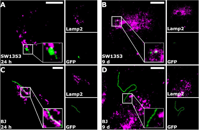
FIGURE 6.
Borrelia burgdorferi did not co-localize with lysosomes. Merged representative images of SW1353 (A,B) and BJ (C,D) cells infected with B. burgdorferi at 24 h and 9-day time points, respectively. Borrelia fluorescence green, lysosomes in magenta, and co-localized pixels appear white. Zoomed images are shown in the white boxes at the bottom right corners of the merged images. As indicated in the merged images, co-localization of B. burgdorferi with lysosomes was not observed. Intermodes threshold was applied to the images before analysis with JaCoP plugin in ImageJ. Scale bars 10 μm.

