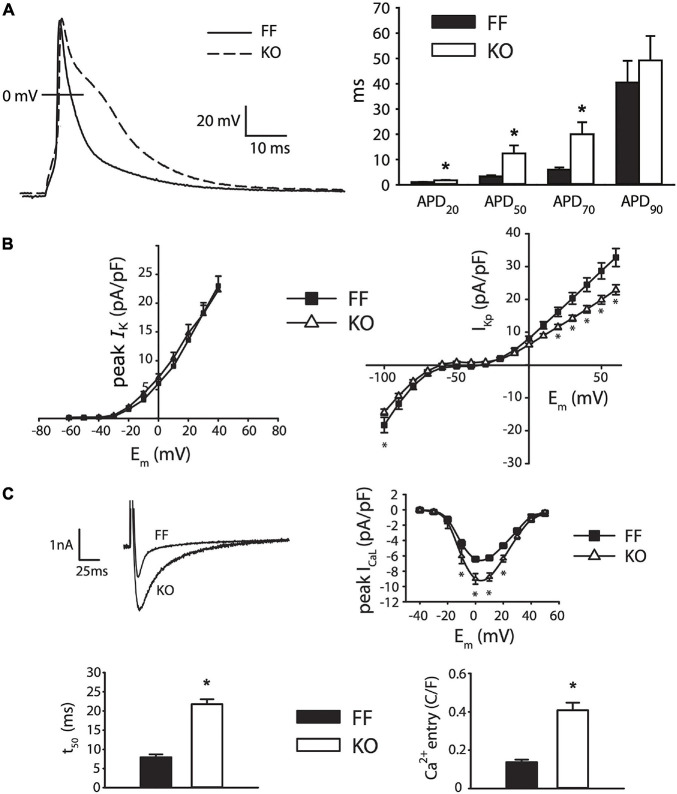
FIGURE 2.
Myocytes from failing SERCA2-KO hearts exhibit prolonged early repolarization due to exaggerated sarcolemmal calcium flux. (A) KO myocytes (n = 10) display marked prolongation of early repolarization compared to FF (n = 11), although this difference is normalized by 90% repolarization. (B) The dominant outward current during early repolarization (Ito), is unaltered in the failing KO myocytes relative to FF (n = 10, both groups; left panel), while slowly inactivating K+ current components (measured as end-pulse, “pedestal” or “plateau,” K+ current – IKp) were slightly suppressed in KO cells (n = 17) relative to FF (n = 20). (C) However, peak ICaL is potentiated and ICaL inactivation is dramatically slowed in KO cells (n = 14) versus FF (n = 12), leading to a marked increase in calcium influx. All panels: *p < 0.05. Data are means ± SEM.

