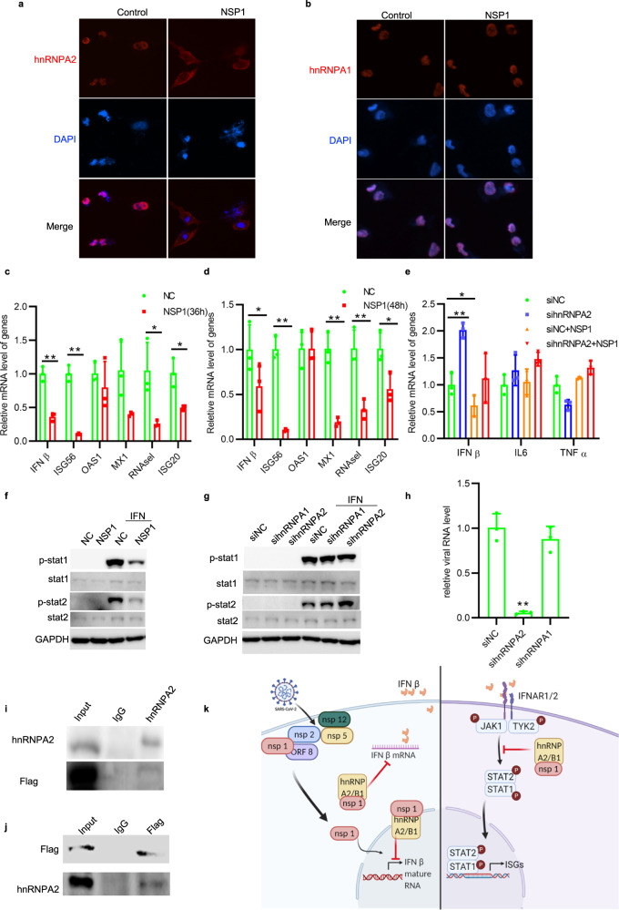
Fig. 1.
a RD cells were ectopically expressed NSP1. After 48 h, hnRNP A2/B1 subcellular location is observed under a fluorescence microscope. hnRNP A2/B1 is indicated as Red. Blue is DAPI. b RD cells were ectopically expressed NSP1 at 48 h. After 48 h, hnRNP A1 subcellular location is observed under a fluorescence microscope. hnRNP A1 is indicated as red. Blue is DAPI. c, d HEK293T cells were transfected with NSP1 for 36 or 48 h. Then mRNA level of IFNß and its downstream ISGs was detected by RT-qPCR, unpaired t test, P < 0.01. e 293T cells were firstly silenced hnRNP A2/B1 by specified siRNA for 24 h, following the ectopic expression of viral protein NSP1 for 48 h. The mRNA levels of IFNß, TNF alpha, and IL-6 in 293T cells were determined by qRT-PCR, unpaired t test, P < 0.05. f HEK293T cells were transfected with control/NSP1 plasmids for 48 h, then treated with/without IFNα (1000 U/ml) for 30 min. Cell lysates were collected for western blot assay. g HEK293T cells were transfected with specific siRNA for 24 h followed by 1000 U/ml of IFNα treatment for 30 min. Cell lysates were collected for western blot assay. h RT-qPCR result of intracellular viral RNA level in RD cells infected with HCoV-OC43 at 72 h post infection, unpaired t test, *P < 0.05. Cells were firstly treated with siNC, sihnRNP A2/B1, and sihnRNP A1 siRNA separately, and then infected with hCov-OC43 after 24 h. i, j 293T cells were ectopically expressed NSP1 at 48 h. Specific antibodies anti-Flag or anti-hnRNP A2/B1 were used for protein immunoprecipitation assay. Normal rabbit or mouse antibodies were used as control. k Scheme (created with BioRender.com.) of SARS-CoV2 NSP1 in regulating host immune response

