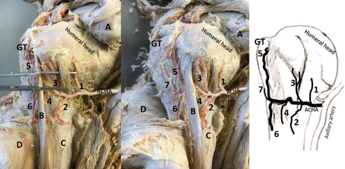
FIGURE 1.

Anterior view of same left proximal shoulder (second image: slightly rotated) with exposure of the branches of the anterior circumflex humeral artery (ACHA; ethics approval: University of Pretoria 70/2017)—most common course, red latex injected. A: reflected pectoralis minor; coracobrachialis and short head of the biceps brachii; B: long head of the biceps brachii tendon; C: latissimus dorsi tendon; D: reflected deltoid; GT: greater tubercle; Pre‐tubercular branches—1: branch that supplies inferior glenohumeral capsule, subscapularis and possible lower part of lesser tubercle; 2: medial descending branch (latissimus dorsi tendon and lower long head of biceps tendon); 3: medial ascending branch (enters medial edge of intertubercular groove); 4: branch in intertubercular groove. Post‐tubercular branches—5: anterolateral (lateral ascending) branch; 6: (lateral) descending branch; 7: possible “transverse branch”
