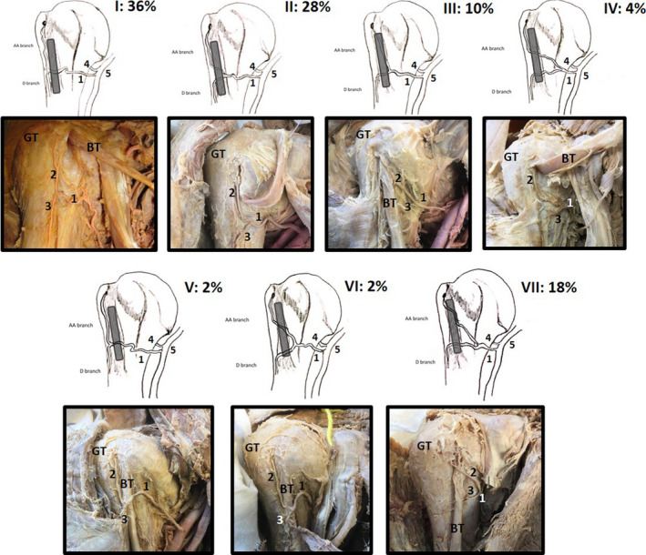
FIGURE 2.

Illustrations of the anterior proximal humerus demonstrating the 7 (I–VIII) identified variations (ethics: University of Pretoria 70/2019) of the ACHA (1), its anterolateral branch (AA branch, 2) and its descending branch (D branch, 3) in relation to the long head of the biceps tendon (BT; shaded in grey). Variations I–IV illustrate the ACHA and branches lying deep to the BT; in variations V and VI, the ACHA and its branches lie anterior. Variation VII shows the anterolateral branch (2) lying anterior to the BT while the descending branch (3) lies deep to the BT (GT: greater tubercle; 4: posterior circumflex humeral artery; 5: axillary artery). All percentages represent the occurrence of these variations in a sample of 50 shoulders
