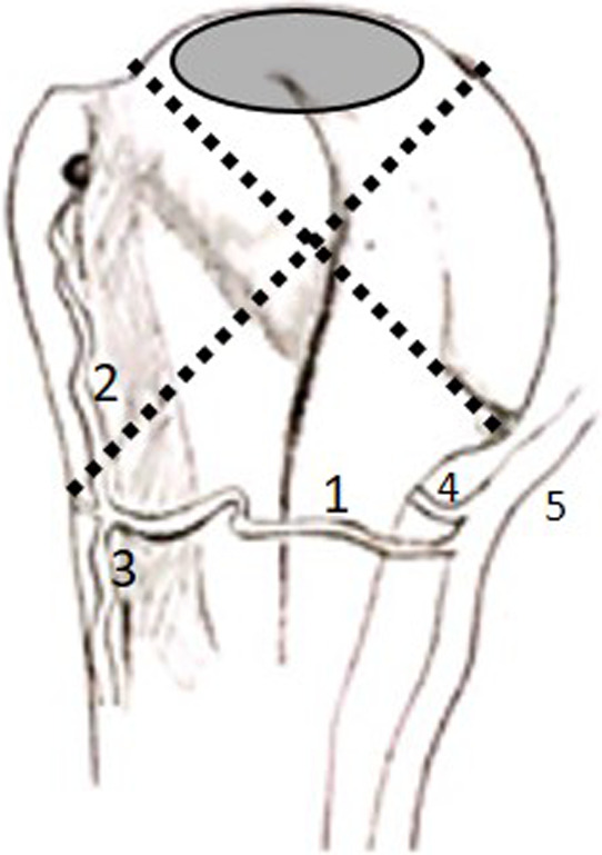
FIGURE 3.

Illustration of the anterior view of the proximal humerus demonstrating the quadrants (dotted lines) outlined by Hettrich et al., (2010) and the poorly vascularized zone (grey oval) identified by Keough et al., (2019) (1: anterior circumflex humeral artery; 2: anterolateral branch; 3: descending branch; 4: posterior circumflex humeral artery; 5: axillary artery)
