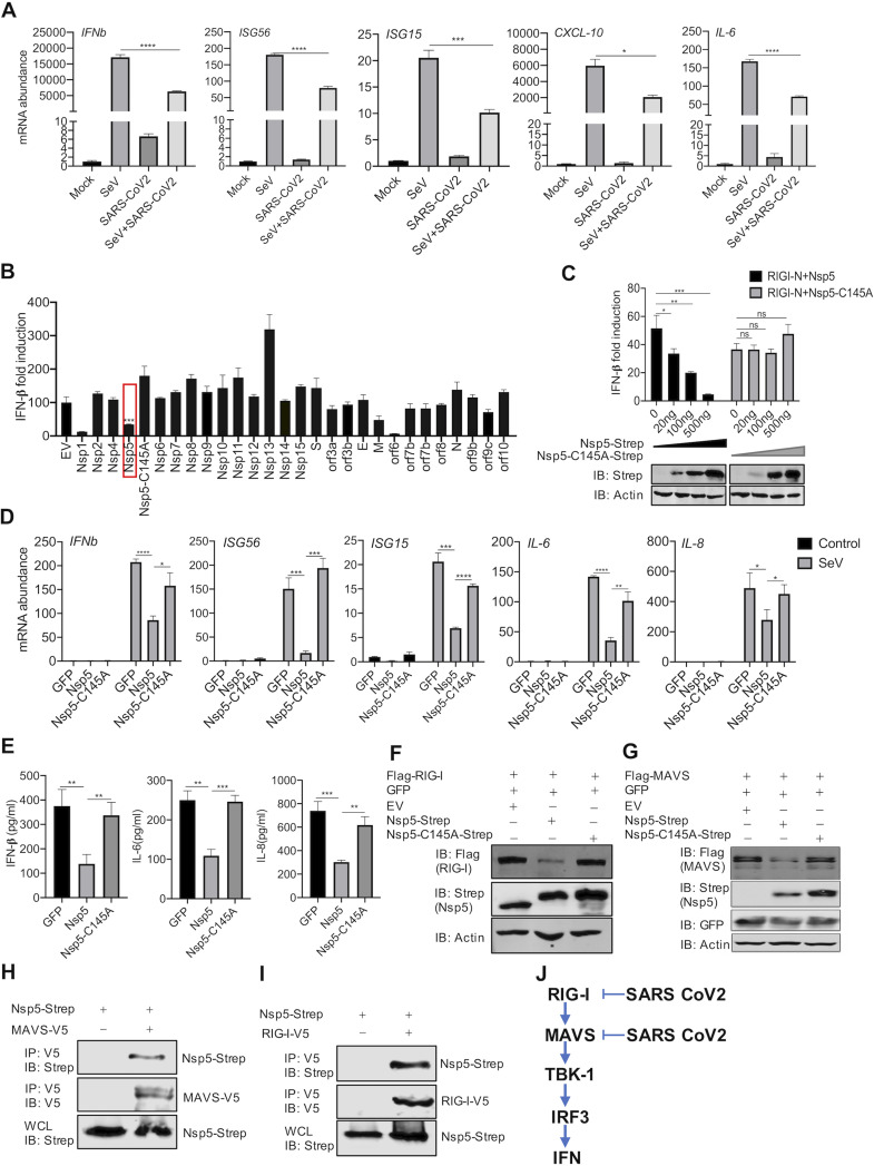
FIG 1.
SARS-CoV-2 Nsp5 targets RIG-I and MAVS to inhibit IFN induction. (A) Calu-3 cells were mock infected or infected with SARS-CoV-2 (MOI = 0.5) for 24 h and superinfected with Sendai virus (SeV; 100 hemagglutinating units [HAU]/ml) for 6 h. The expression of the indicated antiviral genes was analyzed by real-time PCR using total RNA. (B) A screen identified SARS-CoV-2 proteins that inhibit IFN induction by the ectopically expressed MAVS in 293T cells. (C) IFN induction in 293T cells stimulated by RIG-I-N was assessed by a reporter assay with wild-type Nsp5 or its enzyme-deficient Nsp5-C145A mutant. IB, immunoblotting. (D, E) Antiviral cytokine gene expression in HCT116 cells stably expressing Nsp5 or the Nsp5-C145A mutant in response to SeV infection (100 HAU/ml) was analyzed by real-time PCR (D) and ELISA (E). (F, G) Immunoblotting analysis of ectopically expressed RIG-I (F) and MAVS (G) with Nsp5 or the Nsp5-C145A mutant. (H, I) Interactions of SARS-CoV-2 Nsp5 with RIG-I (H) or MAVS (I) were analyzed by coimmunoprecipitation in transfected 293T cells. (J) Diagram of the point of inhibition by SARS-CoV-2 of the RIG-I-MAVS pathway. Data are means ± standard deviations (SD). Significance was calculated using Student's two-tailed, unpaired t test. *, P < 0.05; **, P < 0.01; ***, P < 0.001; ****, P < 0.0001; ns, nonsignificant. See also Fig. S1 in the supplemental material.

