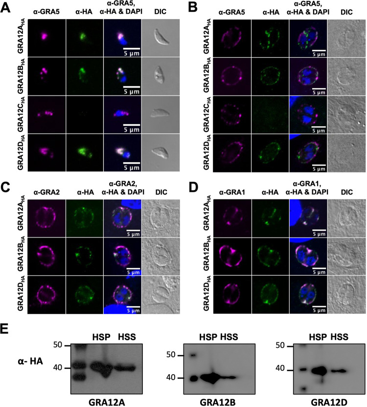
FIG 1.
GRA12-related proteins localize to tachyzoite dense granules, are secreted into the PV, and behave as a soluble and transmembrane PV proteins. (A) Extracellular tachyzoites were stained with anti-HA (α-HA) and anti-GRA5 (α-GRA5) antibody. Tachyzoite nuclei were stained with 4′,6-diamidino-2-phenylindole (DAPI). Tachyzoites were located using differential interference contrast (DIC) microscopy and imaged by confocal microscopy. Number (n) of tachyzoites analyzed (n = 46 for GRA12A, n = 39 for GRA12B, n = 9 for GRA12C, n = 19 for GRA12D). Bars = 5 μm. (B to D) Human foreskin fibroblast (HFF) cells on coverslips were infected with 0.5 MOI tachyzoites of each strain. Vacuoles were located using DIC microscopy, and parasite and host nuclei were stained with DAPI and imaged by confocal microscopy. Vacuoles were stained with anti-HA and anti-GRA5 (B), anti-GRA2 (C), or anti-GRA1 (D). Number (n) of vacuoles analyzed, 6 to 42. Bars = 5 μm. (E) PV proteins were fractionated by ultracentrifugation into high-speed pellet (HSP) and high-speed supernatant (HSS) fractions. Proteins were resolved by electrophoresis, transferred to membranes, and probed with anti-HA rabbit antibody specific for HA-tagged GRA12A, GRA12B, or GRA12D, labeled with goat anti-rabbit IgG coupled to peroxidase, and visualized by chemiluminescence.

