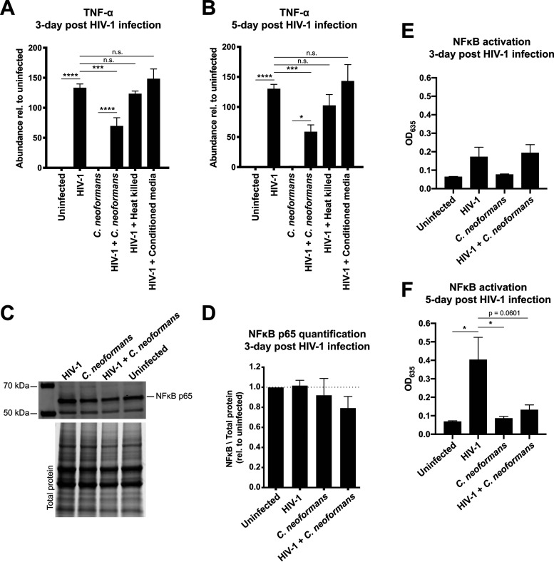
FIG 2.
C. neoformans infection of HIV-1-infected human THP-1 macrophages dampens TNF-α levels and NF-κB signaling. (A and B) Infection for (A) 3 or (B) 5 days with HIV-1 upregulated the TNF-α expression, while C. neoformans infection of HIV-1-infected macrophages dampened the TNF-α transcript abundance. TNF-α abundance was determined using RT-qPCR. Heat-killed cells and conditioned medium did not yield a significant change. (C) Immunoblot analysis probing for the NF-κB subunit p65. The bottom panel shows the total protein as a loading control. (D) Quantification of the p65 bands showed no change in the protein levels. p65 was normalized to total protein and graphed relative to uninfected THP-1 macrophages. (E) NF-κB activity at 3 days post-HIV-1 infection. THP-1 Blue cells were infected with indicated pathogens, and cell culture medium was collected. NF-κB activity was detected using a secreted alkaline phosphatase assay. (F) NF-κB activity at 5 days post-HIV-1 infection. The error bar shows the standard error of the mean (SEM). One-way analysis of variance (ANOVA) with Tukey’s multiple-comparison test was performed to show significance.

