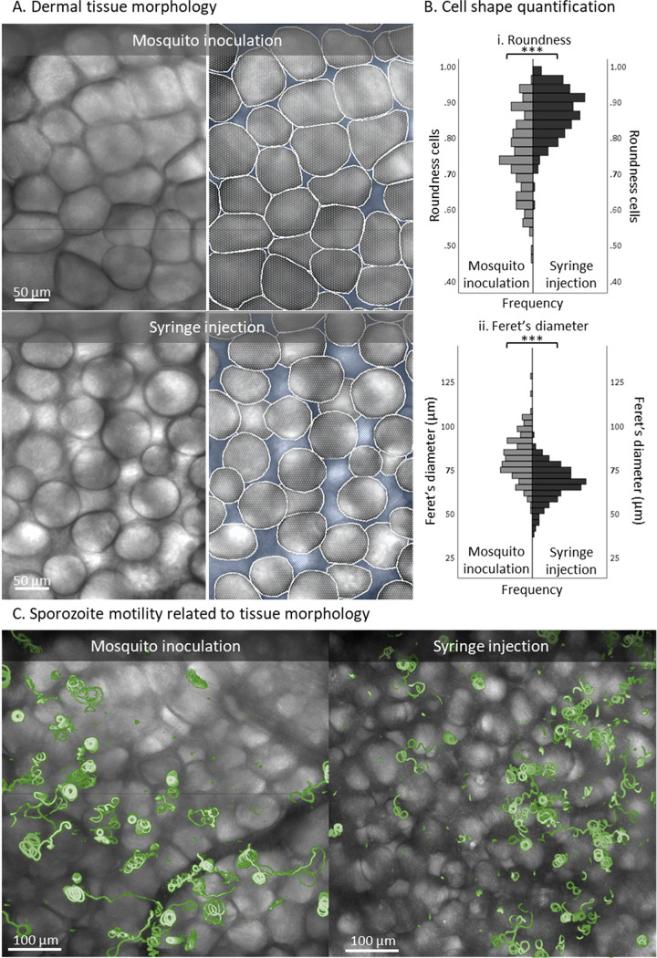
FIG 4.
Magnifications of dermal tissue morphology after sporozoite delivery. (A) Magnification of the tissue morphology of the inoculation site after sporozoite delivery by mosquito and of the injection site after sporozoite delivery by intradermal syringe injection. Based on the bright-field images, the cells (depicted in white) and the interstitial space (depicted in blue) were segmented. (B) Quantification of the cell shapes found after mosquito inoculation (n = 164) and syringe injection (n = 203), using roundness (panel i) and Feret’s diameter (the longest distance between any two points along the cell membrane) (ii) as measures. ***, P < 0.001; independent sample t test. (C) Overview of the dermal site shown as an overlay of a bright-field image and a map of mosquito-inoculated and syringe-injected sporozoite tracks (depicted in green).

