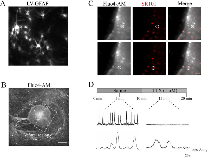
Fig. 1.
SR101-positive glial cells display spontaneous, slow Ca2+ oscillations. A Representative images of GFAP-LV vectors visualizing astrocytes in the ventral-horn of the organotypic spinal slice. Scale bar 50 µm. B Representative fluorescent image at low magnification (4 ×) of a spinal organotypic slice loaded with the calcium dye Fluo4-AM (4 µM). The ventral region (white frame) is identified by the ventral fissure. Scale bar, 500 µm. C CCD-camera snapshots visualize cells located at the border of the ventral region and loaded with Fluo4-AM, in grey (left), and with SR101, in red (middle). Merged images on the right. Scale bars, 50 µm. D Top, representative fluorescent tracing of spontaneous neuronal Ca2+ activity, prior and after 1 µM tetrodotoxin (TTX), recorded from SR101 negative cell (same as in C). Bottom, representative fluorescent tracing of astrocytes Ca2+ oscillations, before and after the application of TTX, recorded from SR101 positive cell (same as in C)

