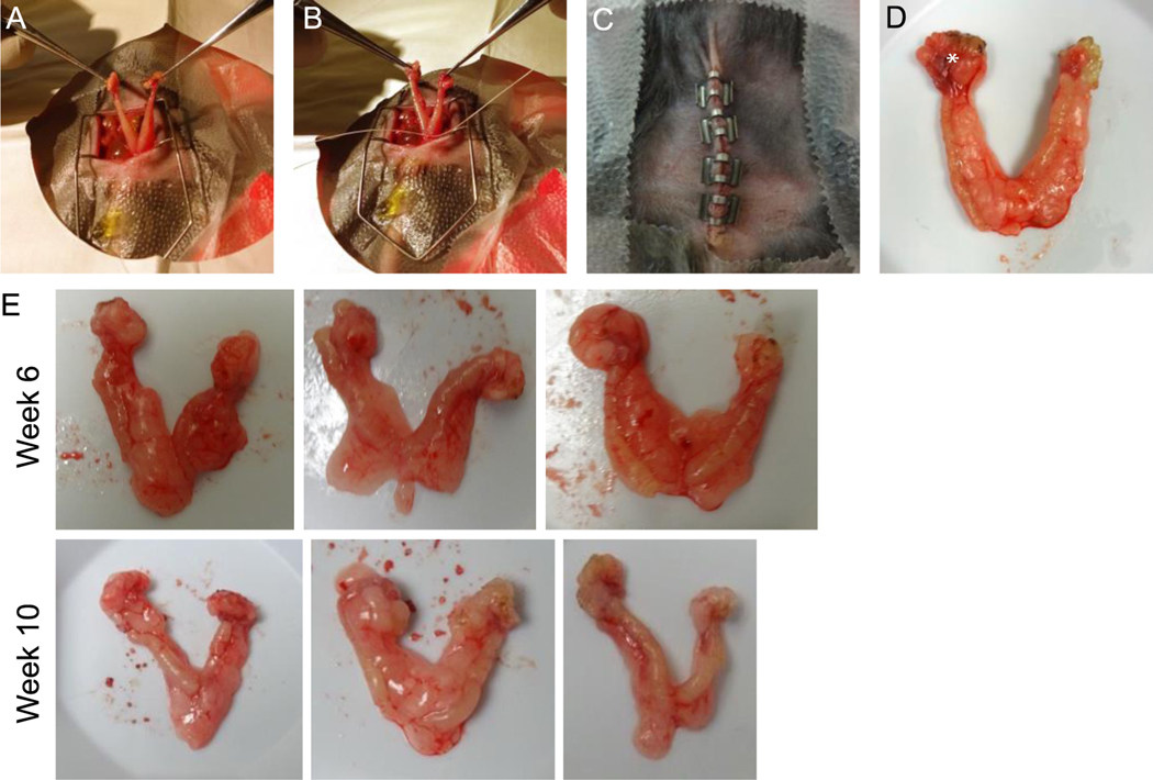
Figure 3. Surgical procedures and surgical specimen after orthotopic injection.
(A) Midline entry into the abdominal cavity with exposure of the bilateral ovaries, fallopian tubes, and uterus. (B) Single suture ligature at the base of the two uterine horns prior to excising the specimen. (C) Closed abdominal cavity with clips inserted at the end of the procedure. (D) Surgical specimen after surgical cytoreduction showing enlarged tumor-bearing right ovary (*). (E) 3 Representative images of surgical specimens from 6- or 10- week cytoreduction. Images are representative of 60 mice from 3 independent experiments.

