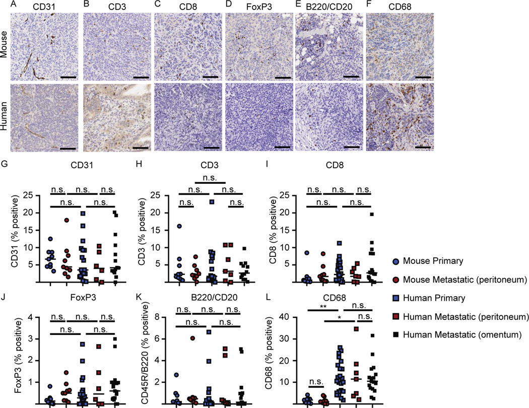
Figure 5. Immunohistochemistry of primary and metastatic tumors.
IHC staining was performed on primary or metastatic tumors from peritoneal surfaces from mice that underwent orthotopic tumor injection but no cytoreductive surgery, and all samples were obtained when mice reached a terminal endpoint necessitating euthanasia. Representative images of (A) CD31, (B) CD3, (C) CD8, (D) FoxP3, (E) B220/CD20, and (F) CD68 from mouse or human samples, respectively. Percent cells positive for the defined molecule from primary and metastatic mouse and human tumors (G) CD31, (H) CD3, (I) CD8, (J) FoxP3, (K) B220/CD20, and (L) CD68, respectively. Scale bars represent 100 μm. Images (A-F) are representative of 9 primary mouse tumors and 19–24 primary human tumors. Data (G-L) from 9 mice per group, 19–24 patient samples per group. Statistical analysis was performed using one way ANOVA for multiple comparisons.

