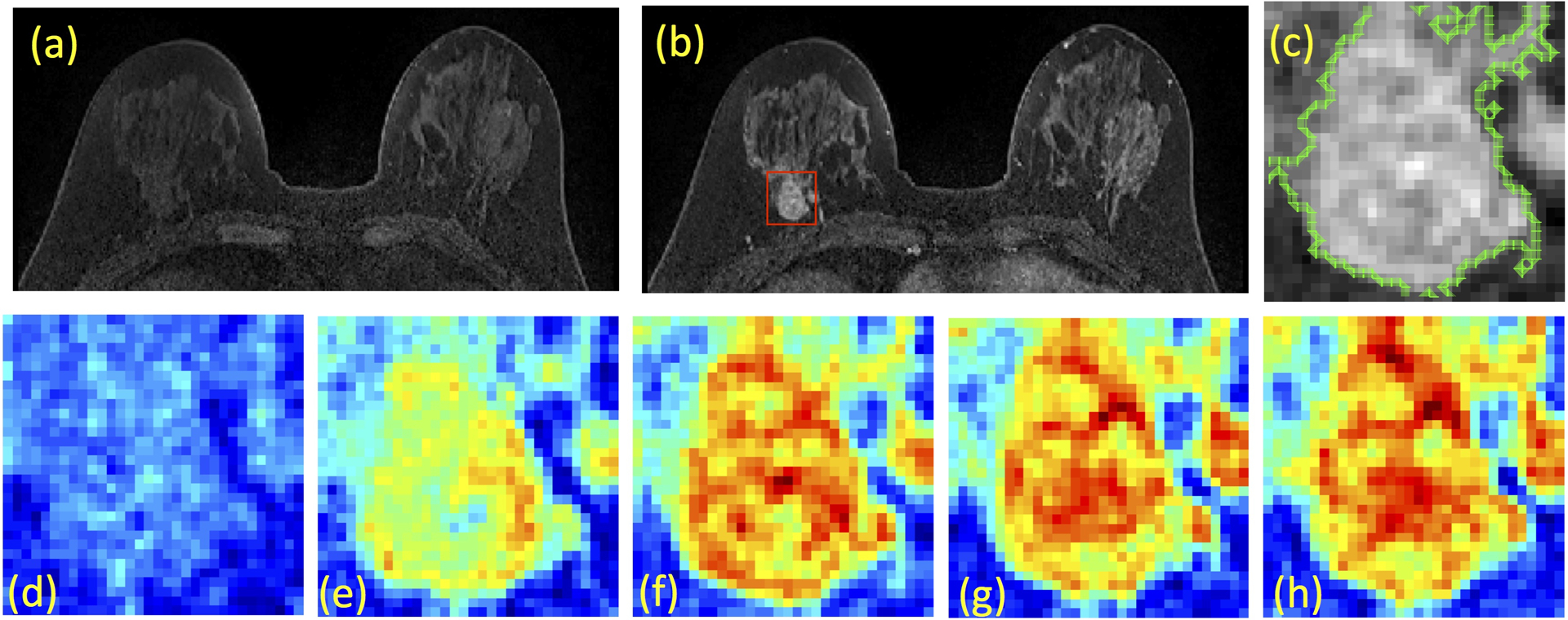
Figure 2:

A case example from a 48-year-old woman with Hormonal-positive and HER2-negative breast cancer in the right breast. (a) Pre-contrast image, (b) Post-contrast image, (c) The zoom-in image of the lesion with outlined tumor boundary obtained from segmentation. The square box is centered at the centroid of the tumor. (d-h) Color-coded DCE images at 5 time frames, one pre-contrast and 4 post-contrast, normalized using the same signal intensity scales. Although this patient has moderate breast parenchymal enhancement (BPE), the lesion boundary is clearly visible and can be segmented with computer algorithms.
