. 2021 Aug 12;21(21):4104–4117. doi: 10.1039/d1lc00347j

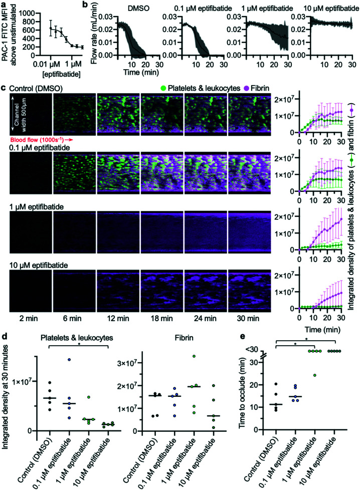
Fig. 5. Concentration-dependent inhibition of occlusive thrombosis by eptifibatide (a) whole blood flow cytometry shows concentration-dependent inhibition by eptifibatide of antibody PAC-1 binding to activated integrin αIIbβ3 (n = 5). (b) Changes in the concentration of eptifibatide were reflected in flow rate data collected from the developed microfluidic assay. (c) Platelets (and leukocytes) and fibrin were visualised using confocal microscopy over a 30 min time-course. The images are representative of n = 5 donors (matched with flow cytometry data). Mean platelet and fibrin accumulation ± S.D. is shown in the graphs (n = 5). (d) The platelet and fibrin integrated density values at 30 min. Treatment with 10 μM eptifibatide had a significant effect on platelet accumulation (P = 0.02(*)). (e) Time to occlude for each experiment is shown in the graph. Treating with 10 μM and 1 μM eptifibatide had a significant effect on the time to occlude compared to control (10 μM eptifibatide P = 0.0197(*); 1 μM eptifibatide P = 0.0423(*)).

Fig. 5