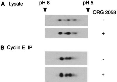
FIG. 2.
Two-dimensional electrophoretic mobility of p27Kip1. T-47D cells were treated and cellular extracts were collected as described for Fig. 1. (A) Extracts from control and treated cells were precipitated, resuspended in IPG buffer, separated by two-dimensional electrophoresis, and Western blotted with a p27Kip1 antibody. (B) Cyclin E-associated p27Kip1 was immunoprecipitated from control and treated lysates using a cyclin E antibody bound to protein A-Sepharose beads. Immunoprecipitated proteins were resuspended in IPG buffer, separated, and analyzed as for panel A. Approximate isoelectric points are indicated.

