
FIG 8.
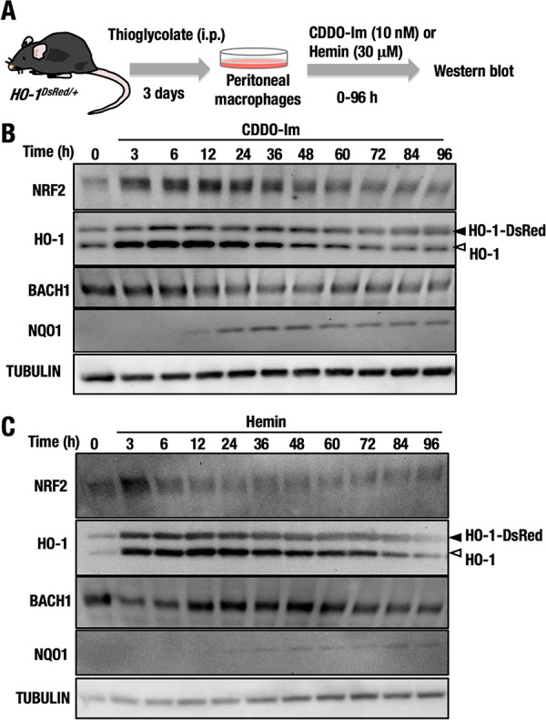
NRF2 and BACH1 expression over time in HO-1DsRed/+ macrophages treated with CDDO-Im and hemin. (A) Thioglycolate-induced peritoneal macrophages were isolated from HO-1DsRed/+ mice, and the macrophages were treated with 10 nM CDDO-Im or 30 μM hemin for 0 to 96 h. (B and C) The protein levels of NRF2, HO-1, BACH1, NQO1, and tubulin in macrophages treated with CDDO-Im (B) or hemin (C) for 0 to 96 h were examined by Western blot analysis. Open and closed arrowheads indicate HO-1-DsRed and endogenous HO-1, respectively.