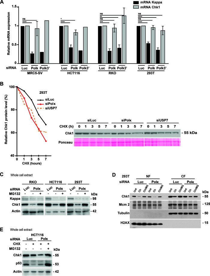
FIG 6.
Pol κ protects Chk1 from degradation. (A) Transcript level analysis of Pol κ and Chk1 genes by RT-qPCR in MRC5-SV, HCT116, RKO, and 293T cells transfected with control siRNA (Luc), siRNA targeting the 3′ UTR of Pol κ (Polκ3′) or the coding sequence of Pol κ. Relative expressions were normalized to the siLuc condition. Values are the means (±SEM) of medians of independent experiments. *, P < 0.05; **, P < 0.01; ***, P < 0.001; ****, P < 0.0001; t test). (B) Western Blot analysis of Chk1 in 293T cells transfected with the indicated siRNA for 48 h and treated with 50 μg/ml of cycloheximide (CHX). Lysates were prepared at the indicated times after cycloheximide addition. Quantification of the Chk1 protein level is shown graphically (left). Ponceau is shown as a protein-loading control. (C) Western blot analysis of Chk1 in RKO, HCT116, and 293T cells. At 48 h after transfection with a control siRNA (Luc) or Pol κ siRNA (Polκ), cells were treated or not with MG132 (20 μM) for 4 h just after transfection and then 6 h before to harvest. Actin is shown as a protein-loading control. (D) Western blot analysis of NF and CF fractions from 293T cells. At 48 h after transfection with a control (Luc) or Polκ siRNA, cells were treated with 50 μg/ml of cycloheximide alone (Ch) or in combination with MG132 (20 μM, ch/MG) for 8 h. (E) Western Blot analysis of Chk1 in HCT116 cells extracts. At 48 h after transfection with indicated siRNA, cells were treated with 50 μg/ml of cycloheximide (CHX) in addition or not to MG132 (20 μM) for 5 h. Actin is shown as a protein-loading control.

